乳腺癌转移至颌骨5例报告
口腔颌面外科杂志 1999年第4期第9卷 经验交流
作者:叶惟熊, Monica B. Zieper, Eugene O.Kelley
单位:School of Dentistry, Oregon Health Sciences University, Portland, Oregon, USA.
BREAST CARCINOMAS METASTATIC TO THE JAWS REPORT OF FIVE CASES
Wei-Yung Yih, Monica B. Zieper, Eugene O. Kelley
中图分类号:R739.8 文献标识码:B 文章编号:1005-4979(1999)04-342-05
Breast carcinomas metastatic to the jaws report of five cases
Metastases of malignant tumors is uncommonly seen in the oral cavity. Reported frequencies range from 1%[1~4] to 8%[5]. Metastases occur in the soft as well as in the osseous tissue of the upper and lower jaw. Bone involvement appears to be more common than soft tissue. However, the criteria used in the literature for the acceptance of a case as a definite metastasis have varied. The criteria of a metastatic case suggested by Causen and Paulsen are as follows: a) a proven rimary tumor with histologic confirmation and, whenever possible, with radiographic supportive evidence; b) maxillary, mandibular or mucosal metastasis with histologic confirmation and with radiographic evidence if the bone is involved; c) histologic correlation of the metastatic oral lesion with the primary lesion; d) when the primary lesion is anatomically near the metastasis, direct extension had to be ruled out by a wide, clear margin around the primary site with no tumor tissue present between the two foci[6]. Causen and Paulsen found 92 carcinomas metastatic to the jaws from 1884 to 1961 which fulfilled the aforementioned criteria[6]. McMillan and Edward, in a survey of the literature from 1966 to 1973, found an additional 166 cases[7]. In a review of 115 cases metastatic to the jaws, Batsakis found that the most common site of primary tumor was the breast[2].
Breast carcinomas metastatic to the jaws usually occurred after other known metastases. Early or solitary metastasis in the jaws may not exhibit typical manifestations of a metastatic tumor. This may cause difficulty in diagnosis and differential diagnosis of the disease. The purpose of this article is to report five additional cases of breast carcinoma metastatic to the jaws. We discuss the signs and symptoms of the disease, existence of problems of diagnosis and differential diagnosis. A correct diagnosis is not only essential for the proper management of such lesions, preventing inappropriate treatment, but also may prolong the life of a patient.
Materials
A total of 21 cases of metastatic tumors in the jaws from 1963~1998 were found in the oral tumor registry of Oregon Health Sciences University School of Dentistry. Five of these 21 cases were of breast carcinoma origin. All cases were female. The ages when their metastatic tumor was discovered ranged from 31~63 years. Four cases were in the mandible (three cases in the left and one case in the right) and one case was located in the anterior maxilla (the apex of the right central incisor). Radiologically, all lesions exhibited an ill-defined osteolytic feature(Fig.1a and Fig.2a). Histopathologically, all cases were adenocarcinoma (Fig 1b, Fig 2b). The interval between the primary tumor diagnosed and the appearance of secondary deposit was from 3 months up to 8 years (Table 1). One case exhibited only toothache which led to the discovery of the metastatic lesion. The rest(4 cases in the mandible) manifested swelling, pain, paresthesia, ulceration, secondary infection and bone exposure.
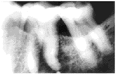
Figure 1a Case 3
Radiograph showing an ill-defined radiolucency associated with vital left lower first molar.
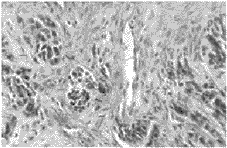
Figure 1b Case 3
Photomicrograph showing an adenocarcinoma.Hematoxylin and eosin Original magnification ×400
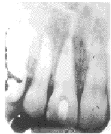
Figure 2a Case 4
Radiograph showing an ill-defined radiolucency associated with a vital right upper central incisor.

Figure 2b Case 4
Photomicrograph showing an adenocarcinoma.Hematoxylin and eosin Original magnification ×400.
Table 1 Breast carcinomas metastatic to the jaws
| Cases |
Sex |
Age |
Location |
Histopathological
Diagnosis |
Interval between Primary
and Secondary Tumor |
Other Regions
of Metastasis |
| 1 |
Female |
44 |
right mandible |
adenocarcinoma
-lobular type |
15 months |
rib |
| 2 |
Female |
48 |
left mandible |
adenocarcinoma
-lobular type |
unknown |
unknown |
| 3 |
Female |
63 |
left mandible |
adenocarcinoma
-lobular type |
8 years |
other bone lesions |
| 4 |
Female |
31 |
anterior maxilla |
adenocarcinoma
-lobular type |
3 months |
lungs |
| 5 |
Female |
51 |
left mandible |
adenocarcinoma
-lobular type with
pagetoid alteration |
1 year |
brain
lung |
Discussion
It is estimated that 1% of malignant neoplasms metastasize to the jaws. Little is known about the pathophysiological mechanisms of this metastatic process. Batsakis propsed that, for a metastatic lesion to become established in bone, there is at least a partial dependence upon the presence of red marrow[2]. Here, thin-walled vascular channels provide a suitable site for lodgment and proliferation of neoplastic emboli. In accordance with this, secondary lesions should most often be found in the molar region. The infrequency of mandibular inolvement may be explained by lack of red marrow in the age group where carcinoma is most prevalent. Sasaki et al conducted an experimental bone metastasis model of the oral and maxillofacial region[8]. They infected human breast cancer (MDA-MB-231) cells into the left heart ventricle of 4 week old female nude mice leading to the development of metastases in the jaws.
Metastases to the jaws are almost always from regions below the clavicle[2].
From several literature review reports, breast carcinomas is the most common origin of metastasis into the jaws[2,7,9,10]. In our oral tumor registry, 5 out of 21 cases of metastatic tumors in the jaws were from breast carcinoma. The metastatic foci can occur in the maxilla and the mandible[2] or bilateral mandible[7] simulataneously. The lesions involved the mandible more commonly than the maxilla with a ratio of 5∶1[1]. In our case, the ratio was 4∶1.
The interval between the diagnosis of the primary tumor and that of metastasis in the jaws varied. In our cases, it was from 3 months to 8 years. Bucin et al reported a case where metastasis developed 15 years after the breast carcinoma was first diagnosed[1]. Therefore, five year cure rate is not always significant and long term follow-up is necessary. Symptoms of metastasis when present are pain and swelling of the affected area. Pain may precede radiological evidence of the lesion by as long as 15 months[2] When the mandible is involved, the earliest sign may be anesthesia over the peripheral distribution of the inferior alveolar nerve on the affected side. Loosening of teeth without evidence of periodontitis or pathological fracture has also been stressed as a possible early sign. Pruckmayer et al demonstrated that toothache was the only sign in their four cases of metastatic breast caricinoma in the jaws[4]. In case 4 of our series, a 31 year-old female with a history of spontaneous pain in the area of the right upper central incisor, radiography showed an ill defined radiolucent area adjacent to the apex of the right upper central incisor (Fig 1a). A 1.5 cm slightly raised magenta-colored area on the facial gingiva over the root of the right upper central incisor was seen. The tooth was sensitive to cold. The clinical manifestaion of this case resembled a periapical inflammatory process except the tooth was sensitive to cold[11]. The patient had breast carcinoma which was treated surgically. She had just completed a course of chemotherapy. Her endodontist curetted the apical lesion. Histopathologic examination of the lesion showed an adenocarcinoma (Fig.1b). Chext x-ray revealed both lungs also had metastatic foci. She died 3 months after the metastatic lesions were discovered.
Breast carcinoma in the jaws usually exhibits an ill-defined osteolytic focus radiographically. However, some breast carcinomas may stimulated new bone formation in the metastatic site resulting in radiopaque or mixed radiolucency with opacity[12,13]. These findings may cause difficulty in the differentiated diagnosis radiographically.
Diagnosis mainly relies on histolopathologic findings. If the metastatic tumor is well-differentiated and closely resembles a carcinoma of a specific site, the pathologist can say with reasonable certainty that a given metastatic tumor comes for a specific primary site. However, more often, metastatic carcinomas are poorly-differentiated and histopathologic study of the metastatic deposit gives little clue as to the primary site of the tumor. In our cases, all showed an adenocarcinoma, lobular type. Less differentiated adenocarcinoma was present in case 5, in which neoplastic cells invaded overlying epithelium resulting in an unusual pagetoid appearance (Fig. 3). Between 20%~30% of the grou had jaw metastases as the first indication of harboring a malignant neoplasm. In such instances, seeking a primary neoplasm based upon the frequencies of metastases is practical. Batsakis tabulated the site of origin of 115 cases of primary neoplasms that metastasized to the jaws as:breast 30.4%, kidney 15.6^, lung 14.8%, colon and rectum 7.8%, prostate gland 7%, thyroid gland 6.1%, stomach 5.2%, skin (melanoma) 4.4%, testes 2.6%, and the rest were less than 1%[2]. In our 21 cases of metastatic neoplasms in the jaws, the primary tumors, in order of decreasing frequency, were as follows: breast, lung, skin, colon, stomach, thyroid, and prostate gland. One case was of unknown origin. McDaniel et al found that, of 32 histologically verified cases metastatic to the jaws, the origin of the primary neoplasms could not be located in 9 of the cases[14].

Figure 3 Case 5
Photomicrograph showing an adenocarcinoma (hollow arrow) with pagetoid alteration(solid arrow).Hematoxylin and eosin Original magnification ×400.
Although the diagnosis of metastatic carcinoma can usually be determined by histopathological examination, the final diagnosis depends mostly on a careful medical history and complete physical examination with appropriate laboratory studies[13]. Patients who present with carcinomatous metastases in the jaw have a grave prognosis. About 70% of the patients die within one year of the diagnosis of such a metastasis[2,4,5]. Treatment is chiefly palliative with the intention of providing relief from pain and prolonging life. Resection of the mandible is too debilitating a procedure and does not significantly prolong the life of the patient. Radiation therapy gives variable results and is dependent on the type and responsiveness of the neoplasm.
The two major types of therapy for disseminated breast carcnoma are use of hormonal and cytotoxic agents. Patients whose tumor is positive for estrogen and progesterone receptor proteins (RP) are usually treated with hormonal therapy (anti-estrogen-tamoxifen) as first line therapy. Patients with RP-negative tumors, or those originally RP-positive who have become refractory to endocrine therapy, require chemotherapy with cytotoxic agents. Resposes to these therapies usually last a medium of 6 to 18 months and responders have a significantly prolonged survival compared to non-responders[15]. In patients receiving estrogen replacement therapy who develop metastatic breast carcinoma, with drawel of estrogen replacement therapy alone is a therapeutic option. This often results in regression of metastatic disease[16].
References
[1] Bucin E, Andréasson L, Björlin G. Metastases in the oral cavity: Case report. Int J Oral Surgery, 1982,11:321.
[2] Batsakis JG. Tumors of head and neck: Clinical and pathologic considerations 2nd ed. Baltimore: Williams & Wilkins, 1979:240.
[3] O'Carroll MK, Krolls SO, Mosca NG. Metastatic carcinoma to the mandible: Report of two cases. Oral Surg Oral Med Oral Pathol, 1993,76:368.
[4] Pruckmayer M, Glaser C, Marosi C, Leitha T. Mandibular pain as the leading clinical symptom for metastatic disease: Nine cases and review of the literature. Ann of Oncol, 1998, 9:559.
[5] Oikarinen VJ, Calonius PEP, Saino P. Metastatic tumors to the oral region(1) An analysis of cases in the literature. Proc Finn Dent Soc, 1975,71:58.
[6] Clausen F, Paulsen H. Metastatic carcinoma to the jaws. Acta Pathol Microbiol Scand, 1964,57:361.
[7] McMillan MD, Edwards JL. Bilateral mandibular metastases. Oral Surg Oral Med Oral Pathol, 1975,39:959.
[8] Sasaki A, Yoneda T, Terakodo N, et al. Experimental bone metastasis model of the oral and maxillofacial region. Anticancer Res, 1998,18(3A):1579.
[9] Epker BN, Merrill RG, Henny FA. Breast adenocarcinoma metastatic to the mandible: Report of seven cases. Oral Surg Oral Med Oral Pathol, 1965,20:350.
[10] Meyer I, Shklar G. Malignant tumors metastatic to mouth and jaws. Oral Surg Oral Med Oral Pathol, 1965,20:350.
[11] Yih WY. Zebra XI Part I and Part II. J Endodon, 1992,18(6):307 & 18(7):355.
[12] Thompson CC, Bartley MH, Wooley LH. Metastatic tumors of the head and neck: A 20 year oral tumor registry report. J Oral Med, 1986,41:175.
[13] Neville BW, Damm DD, Allen CM, Bouquot JE. Oral and Maxillofacial Pathology 1st ed. Philadelphia: WB Saunders Company, 1995:489.
[14] McDaniel RK, Luna MA, Stimson PG. Metastatic tumors in the jaws. Oral Surg Oral Med Oral Pathol, 1971,31:380.
[15] Wyngaarden JB, Smith LH Jr. Cecil textbook of medicine 18th ed. Philadelphia: WB Saunders Company, 1988:1456.
[16] Dhodapkar MV, Ingle JN, Ahmann DL. Estrogen replacement therapy withdrawal and regression of metastatic breast cancer. Cancer, 1995, 75:43.
收稿日期:1999-01-09